
What is PET/CT?
Positron Emission Tomography/Computerized Tomography (PET/CT) is an imaging test that produces high resolution pictures of the body’s biological functions and anatomic structures. These images show body metabolism and other functions rather than simply the gross anatomy and structure revealed by a standard CT or MRI scan. This is important because functional changes are often present before obvious structural changes in tissues are evident. PET/CT imaging can uncover abnormalities that might otherwise go undetected. For example, PET/CT can determine the presence and extent of tumors unseen by other imaging techniques, or detect Alzheimer’s disease one to two years before the diagnosis would be made with certainty by your primary doctor.
Diseases for which PET/CT is most commonly used:
- Cancer
- Alzheimer’s Disease
- Coronary Artery Disease
- Epilepsy
How accurate is PET/CT?
PET/CT is believed to be the most accurate imaging test available to evaluate lung cancer, colon cancer, breast cancer, melanoma, lymphoma, head and neck cancer, and esophageal cancer. In published research studies, PET has been shown to have an approximately 90% accuracy in many of these cancer types. PET is the most accurate imaging test available to determine the presence of a dementia process such as Alzheimer’s disease. PET is also the most accurate test available to evaluate patients who have had a previous heart attack and are being considered for a procedure to improve blood flow to the injured heart muscle.
PET/CT assists physicians and patients in the management of cancer by:
- Assessing the location and extent of cancer prior to surgical or radiation treatment
- Serving as a test for early detection of recurrent cancer
- Monitoring the effect of treatment non-invasively
- Reducing or eliminating ineffective and unnecessary treatment
- Replacing multiple medical testing procedures with a single exam
Who should not have a PET/CT scan?
- Pregnant women should consult their doctor before having this scan.
- Patients who have severe kidney problems or who are allergic to iodine can have a PET/CT without the I.V. contrast.
- An itching sensation or shortness of breath may indicate an allergic reaction to the contrast. Please let your technologist know if you have any allergic reaction.
How does the procedure work?
- You will be asked to drink approximately 3 cups of oral contrast.
- Shortly after, you’ll be injected with a radioactive sugar isotope solution.
- You will then rest comfortably for approximately 45 minutes while the sugar distributes throughout your body.
- Before you get positioned on the scanner, you will have a chance to visit the restroom and change into a gown.
- You will be scanned for approximately 30 minutes and may receive additional I.V. contrast depending on your physician’s request. Normal reactions when getting I.V. contrast may include flushing, nausea, headache or a salty taste in your mouth.
What should I do to prepare for the procedure?
- On the day of the scan take your regularly prescribed medications with water only.
- Do not eat or drink anything except water for four to six hours before the scan.
- Diabetic patients should schedule their scan so that their last dose of insulin or diabetic medication is at least two hours prior to the procedure.
- Refrain from heavy exercise for 12 hours prior to the scan.
- Drink at least two glasses of water one hour before the scan.
How will I feel after the exam?
You should feel fine. You may resume your normal activities and eat a regular meal immediately following the scan.
How much time should I allow?
Most patients will be in the clinic for approximately two to two and a half hours.
When will I know the results of my PET/CT Scan?
Your referring physician will have the report 24 to 48 hours after the exam.
The PSMA PET scan is a breakthrough diagnosis to locate prostate cancer tumors for more precise treatment
Why choose PSMA PET/CT Scan for Prostate Cancer?
Precisely locating prostate cancer so that it can be treated is extremely challenging. This is because prostate cancer tumors can be found not only in the prostate but also throughout the pelvis and the body in cases where the tumors have migrated.
Effectively pinpointing and eliminating these tumors is important because prostate cancer is the second most common cancer in men and the second leading cause of cancer death in men.
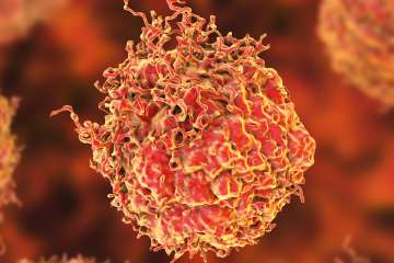
In 2022 alone, an estimated 268,490 cases of prostate cancer will be diagnosed, with about 34,500 deaths
For this reason, UCLA Health and UC San Francisco validated for use in the United States a diagnostic innovation known as the prostate-specific membrane antigen or PSMA PET/CT scan. This scan is approved by the U.S. Food and Drug Administration and it enables more precise detection of prostate cancer for treatment planning and targeted care.
PSMA PET/CT is a better standard of care because it is more accurate
UCLA Health researchers, along with colleagues at UC San Francisco, studied PSMA PET/CT for several years. These studies led to the FDA approval of this imaging technique’s use in the United States.
PSMA PET/CT scans for prostate cancer provide better care for men who are initially diagnosed with prostate cancer or for men who were previously treated but have experienced a recurrence of the cancer. In both cases, PSMA PET/CT is better able to find the extent of this cancer in the body.
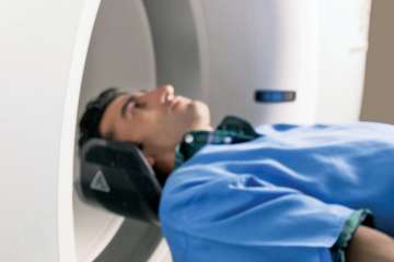
PSMA PET/CT enables more precise detection of prostate cancer
PSMA PET/CT works using a radioactive tracer, called 68Ga-PSMA-11, which is manufactured on site at UCLA Health. Nuclear medicine physicians inject this tracer which attaches itself to proteins known as prostate-specific membrane antigens (PSMA). Because prostate cancer tumors overexpress these proteins on their surface, physicians can pinpoint their location with the tracer.
The current standard of care in prostate cancer imaging is a technique called fluciclovine PET/CT, which involves physicians injecting patients with a synthetic radioactive amino acid.
In their research comparing PSMA and fluciclovine PET/CT, the UCLA and UCSF research teams found that imaging with PSMA PET/CT was able to detect significantly more prostate lesions than fluciclovine PET/CT in men who had undergone a radical prostatectomy but had experienced a recurrence of their cancer.
How to receive a PSMA PET/CT scan and prostate cancer care at UCLA Health
UCLA Health provides comprehensive and customized care for men with prostate cancer. Our physicians use the PSMA PET/CT scan alongside radiation therapy, chemotherapy, surgery, and all other treatment modalities offered to make sure men receive the best possible care – from diagnosis to treatment to follow-up.
To ensure the best treatment possible, UCLA Health’s nuclear medicine physicians, medical oncologists, radiation oncologists, radiologists, urologists and surgeons optimize care for each person receiving treatment. The prostate cancer care at UCLA Health is backed by multidisciplinary tumor boards with physicians from different specialties and subspecialties, along with genetic counselors and representatives from allied health services. Together, these specialists and experts meet once a week to talk about each new cancer patient and the path forward for their specialized treatment.
How to get started with a PSMA PET/CT Scan for prostate cancer
Referrals
UCLA Health welcomes patient referrals for a PSMA PET/CT scan from physicians who are based anywhere in the world. For patients who have questions about gaining a physician’s referral, please contact us.
Connect with us
For an appointment, call us at 310-794-1005. Appointments are taken Monday through Friday, 8 am to 5 pm. Please leave a message after hours.
Internal healthcare providers can e-mail the Nuclear Medicine Clinic at UCLA Health or place an order in CareConnect for individuals who are already patients.
Where treatment happens
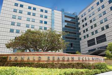
UCLA Health’s PSMA PET/CT scan is performed at the Ronald Reagan UCLA Medical Center on UCLA’s campus in the Westwood area of Los Angeles.
Once the PSMA PET/CT scan is complete, men can receive prostate cancer care at UCLA Health’s hospitals in Westwood and Santa Monica, or at UCLA Health’s Encino medical office. The scan can also be used in conjunction with care at any other hospital where the patient is being treated.
The total time it takes for a PSMA PET/CT scan is about two hours. What will happen is the tracer will be injected by a UCLA nuclear medicine technologist. Then the patient waits one hour before entering the PET/CT scanner for 30 minutes.
What Is Nuclear Medicine?
Nuclear medicine is a safe and painless imaging technology, which uses very small amounts of specially formulated radioactive materials (or tracers) to help diagnose and treat a variety of diseases. Nuclear medicine provides physicians information about medical problems based on how parts of the body function as differing from x-ray, which demonstrates a body’s appearance (or structure).
All nuclear medicine exams involve IV injection, inhalation and/or swallowing of specially formulated compounds (tracers) with imaging at timed intervals. Tracers are attracted to specific organs, bones or tissues in the body, which then are detectable by special types of cameras. Reactions to these tracers are rare. The total amount of radiation a patient receives from a nuclear medicine examination is comparable to that received during a diagnostic x-ray.
Some examinations require special positioning, and most exams take one to two hours to complete though some will take longer.
Nuclear medicine testing is commonly used in children to evaluate bone pain, injuries, infection or kidney and bladder function. Common nuclear medicine applications include:
- Diagnosis and treatment of thyroid conditions
- Cardiac stress tests to analyze blood flow to the heart muscle
- Bone scans for detection of cancer, infection or subtle bone injuries
- Lung scans for blood flow to the lungs and air exchange
- Liver and gall bladder scans